
Research Area 3: Scanning Electrochemical Microscopy (SECM) and Fluorescence Microscopy
The focus of this research area is the development of instrumentations for both fundamental studies of electrochemical reactions and applied biomedical research.
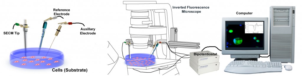
Since its initial development, SECM has been widely used in studying electron transfer mechanisms at interfaces. More recently, it has been shown to be well-suited for cell studies, in particular, when used in tandem with fluorescence microscopy. Specific applications include real time detection of reactive oxygen species in cancer cells and neurotransmitters at synapses.
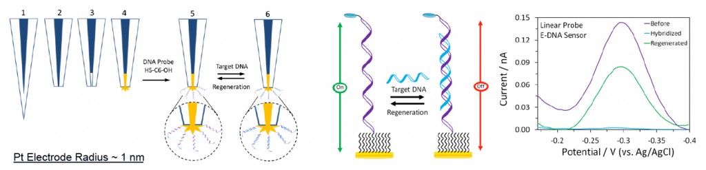
SECM is a versatile electroanalytical technique that is often used with microelectrodes. However, depending on the actual application, miniaturization of the electrode (SECM tip) could be beneficial. We have recently developed a method capable of producing long, sharp and tapered Pt nanoelectrodes using a laser puller. Furthermore, we have successfully fabricated electrochemical DNA sensors on gold-plated recessed Pt nanoelectrodes. These nanoscale sensors are ideal for in vivo detection of disease-relevant targets, including DNA, proteins and small molecules, in living cells.
Recent Publications
– Fabrication of Electrochemical DNA Sensors on Gold-modified Recessed Platinum Nanoelectrodes. Salamifar, S. E., Lai, R. Y., Anal. Chem., 2014, 86,2849-2852.
– Use of Combined Scanning Electrochemical and Fluorescence Microscopy for Detection of Reactive Oxygen Species in Prostate Cancer Cells. Salamifar, S. E.; Lai, R. Y. Anal. Chem., 2013, 85, 9417-9421.